
Mast Cell Association In Hemangiomas and Arteriovenous Vascular Malformations.
Keywords:
Arterio-venousmalformation, Hemangioma, Mast cells
Abstract
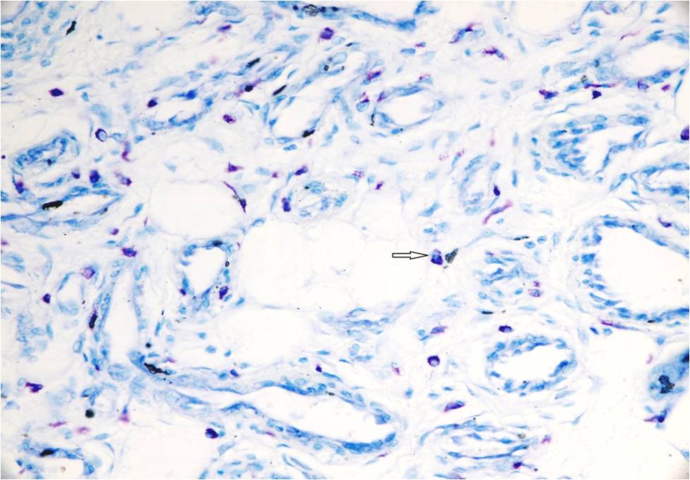
Background: Vascular anomalies, classified as hemangiomas and arteriovenous malformations (AVMs), were analyzed for the presence of mast cells. Hemangiomas in the proliferative phase contained large numbers of mast cells (53.12 ± 27.83 cells/mm2) in comparison with hemangiomas in the involuting phase (11.43 ± 7.9 cells/mm2), and AVM (25.31 ± 27.3 cells/mm2). Considering the fact that hemangiomas are characterized by endothelial proliferation and increased numbers of mast cells, these data raise the possibility that mast cells may have an important role in the formation and/or maintenance of these lesions.Aim: This study aimed at evaluation of presence of mast cells in hemangiomas proliferating, involuting ones and AVMs.Methods: A total of 120 cases of benign vascular lesions were retrieved from 12 years period. A total of 94 cases, where complete clinical details and representative paraffin sections were available, were included in this study. Hematoxylin and eosin (H and E) stain and Mast cell density in all lesions was calculated from toluidine blue stained sections.Results: Mean mast cell density was significantly higher in proliferating hemangiomas (53.12 ± 27.83 cells/mm2) compared to involuting hemangiomas (11.43 ± 7.9 cells/mm2) and AVM (25.31 ± 27.3 cells/mm2)Conclusions: The significantly higher mast cell density seen in proliferating hemangiomas compared with involuting ones, seem to suggest that mast cells play an important role in the natural history of these lesions.References
1. Chang MW. Updated classification of hemangiomas and other vascular anomalies. Lymphat Res Biol 2003;1:259-65.
2. Hand JL, Frieden IJ. Vascular birthmarks of infancy: Resolving nosologic confusion. Am J Med Genet 2002;108:257-64.
3. Adegboyega PA, Qiu S. Hemangioma versus vascular malformation: Presence of nerve bundle is a diagnostic clue for vascular malformation. Arch Pathol Lab Med 2005;129:772-5.
4. Mulliken JB, Glowacki J. Hemangiomas and vascular malformations in infants and children: A classification based on endothelial characteristics. Plast Reconstr Surg 1982;69:412-22.
5. Enjolras O, Mulliken JB. Vascular tumors and vascular malformations (new issues). Adv Dermatol 1997;13:375-423.
6. Girard C, Graham JH, Johnson WC. Arteriovenous hemangioma (arteriovenous shunt). A clinicopathological and histochemical study. J Cutan Pathol 1974;1:73-87.
7. Koutlas IG, Jessurun J. Arteriovenous hemangioma: A clinicopathological and immunohistochemical study. J Cutan Pathol 1994;21:343-9.
8. Burrows PE, Mulliken JB, Fellows KE, Strand RD. Childhood hemangiomas and vascular malformations: Angiographic differentiation. AJR Am J Roentgenol 1983;141:483-8.
9. Carapeto FJ, Garcia-Perez A, Winkelmann RK. Acral arteriovenous tumor. Acta Derm Venereol 1977;57:155-8.
10. Lascano EF. Mast cells in human tumors. Cancer 1958;11:1110-4.
11. Baroni C. On the relationship of mast cells to various soft tissue tumours. Br J Cancer 1964;18:686-91.
12. Glowacki J, Mulliken JB. Mast cells in hemangiomas and vascular malformations. Pediatrics 1982;70:48-51.
13. Azizkhan RG, Azizkhan JC, Zetter BR, Folkman J. Mast cell heparin stimulates migration of capillary endothelial cells in vitro. J Exp Med 1980;152:931-44.
Published
2015-05-10
Issue
Section
Original Article
Authors who publish with this journal agree to the following terms:
- Authors retain copyright and grant the journal right of first publication with the work simultaneously licensed under a Creative Commons Attribution License that allows others to share the work with an acknowledgement of the work's authorship and initial publication in this journal.
- Authors are able to enter into separate, additional contractual arrangements for the non-exclusive distribution of the journal's published version of the work (e.g., post it to an institutional repository or publish it in a book), with an acknowledgement of its initial publication in this journal.
- Authors are permitted and encouraged to post their work online (e.g., in institutional repositories or on their website) prior to and during the submission process, as it can lead to productive exchanges, as well as earlier and greater citation of published work (See The Effect of Open Access at http://opcit.eprints.org/oacitation-biblio.html).

