
Toluidine blue stain and crystal violet stain versus H&E stain in the Diagnosis of Hirschsprung’s Disease: A Study in Sulaimani City in Kurdistan/Iraq
Keywords:
Hirschsprung’s disease, Ganglion cells, Cresyl violet, Toluidine blue, Mast cells
Abstract
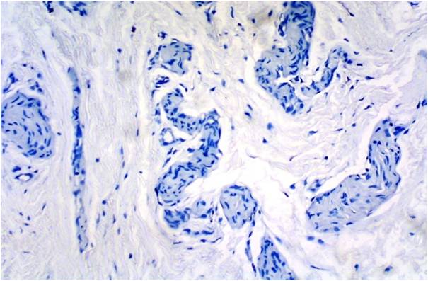
Background Hirschsprung’s Disease (HD) is a congenital disorder of the colon in which certain nerve cells, known as ganglion cells are absent.Setting and Design: To demonstrate the efficacy of Cresyl Violet and Toluidine blue (Tb) special stains in the identification of ganglion cells in suspected Hirschsprung’s disease and to find other adjuvant histological criteria for the diagnosis. Method: In Sulaimani Teaching Hospital and Pediatric teaching hospital in Sulaimani Governorate/ Kurdistan-Iraq a total of fifty non selected cases biopsied for suspected HD were stained with hematoxylin and eosin (H&E) stain and divided into two groups: HD and-non-HD. All cases then should be stained with Tb special stain to identify ganglion cells and to count mast cells in the submucosa. Cases were stained with Cresyl Violet special stain to identify ganglion cells. H&E- and Tb-stained sections were examined for the presence or absence of hypertrophic nerve fibers in the submucosa. Results: Both Cresyl violet and Tb stains were superior to H&E in the identification of ganglion cells with no statistically significant difference between the two stains. Mast cell count in the submucosa has no important effect on diagnosis while nerve bundle hypertrophy was found to be associated with absence of ganglion cells in Hirschsprung disease.Conclusions: Toluidine blue and/or Cresyl violet stains should to be used as the routine stain to highlight ganglion cells in suspected Hirschsprung’s disease cases. Submucosal nerve bundle hypertrophy has to be assessed as an adjuvant histolological criterion.References
1. Worman S, Ganiats TG. Hirschsprung's disease: a cause of chronic constipation in children. Am Fam Physician.1995; 51: 487–94.
2. Hirschsprung's Disease and Allied Disorders. Berlin: Springer. 2007. ISBN 3-540-33934-5.
3. Hirschsprung, H. "Stuhlträgheit Neugeborener in Folge von Dilatation und Hypertrophie des Colons". Jahrbuch für Kinderheilkunde und physische Erziehung(Berlin).1888; 27: 1–7.
4. Kays DW. Surgical conditions of the neonatal intestinal tract. Clinics in Perinatology. 1996; 23 (2): 353–75.
5. Suita S, Taguchi T, Ieiri S, Nakatsuji T. Hirschsprung's disease in Japan: analysis of 3852 patients based on a nationwide survey in 30 years. Journal of Pediatric Surgery. 2005; 40 (1): 197–201; discussion 201–2.
6. Goldman, Lee. Goldman's Cecil Medicine (24th ed.). Philadelphia: Elsevier Saunders. pp. 867. ISBN 1437727883.
7. Lukman O. Abdur-Rahman, Brian H. Cameron. Hirschsprung's Disease in Africa in the 21st Century. Surgery in Africa Monthly Review. Hamilton, Canada. January. 2011 http://ptolemy.library.utoronto.ca/sites/default/files/reviews/2011/January%20-%20Hirschsprung's%20Disease.pdf
8. Kiernan JA. Nissl: the man, the stain and the substance. Microscopy Today. 2005;13(6):50.
9. Bear MF, Connors BW, Paradiso MA. Neuroscience: Exploring the Brain, 3rd ed. Philadelphia: Lippincott,Williams & Wilkins, 2007, pp.31-34;94-98.
10. Rosai J. Large Bowel Disease. In: Ackerman’s Surgical Pathology. Philadelphia: Mosby; 2004. p. 777-9.
11. Martucciello G. Hirschsprung’s disease, one of the most difficult diagnoses in pediatric surgery: a review of the problems from clinical practice to the bench. Eur J Pediatr Surg. 2008;18:140-9.
12. Chen F, Winston JH 3rd, Jain SK, Frankel WL. Hirschsprung’s Disease in a young adult: report of a case and review of the literature. Ann Diagn Pathol. 2006;10:347-51.
13. Petras R. Hirshsprung’s disease. In: Sternberg, SS. Diagnostic surgical pathology, Williams and Wilkins; Philadelphia: 2004. p. 1390-1.
14. STAINING MANUAL - NERVE TISSUE http://library.med.utah.edu/WebPath/HISTHTML/MANUALS/NISSL.PDF
15. Tanisha N. Neely, Diana Harrington. MediaLab course, Histology Special Stains: Nervous Tissue. http://www.medialabinc.net/spg538507/luxol fast_blue_lfb
16. Arline d. Deitch, Montrose J Moses. The Nissl substance of living and fixed spinal ganglion cells. j. Biophyslc. and Biochek. Cytol., 1957;3: 449-455
17. Gokul Sridharan, Akhil A Shankar. Toluidine blue: A review of its chemistry and clinical utility. J Oral Maxillofac Pathol. 2012;16: 251–255.
18. John A. Kiernan. Staining Sections of the Central Nervous System. Canada. 2012: 62
19. Toluidin blue stain for histology. http://reactiveshops.com/index.php?route=product/product&product_id=97
20. Kumar GL, Kiernan JA. 2nd ed. Dako North America, California: 2010. Education guide: Special stains and H & E.
21. Yadav AK, Mishra K, Mohta A, Agarwal S. Hirschsprung’s disease: Is there a relationship between mast cells and nerve fibers? World J Gastroenterol. 2009 March 28; 15(12): 1493–8.
22. Kobayashi H, Yamataka A, Fujimoto T, Lane GJ, Miyano T. Mast cells and gut nerve development: implications for Hirschsprung's disease and intestinal neuronal dysplasia. J Pediatr Surg. 1999;34(4):543-8.
23. M. Canil, K. Meir, G. Jevon, T. Sturby, S. Moerike, A. Gomez. Toluidine Blue Staining Is Superior to H&E Staining for the Identification of Ganglion Cells in Frozen Rectal Biopsies. Technical bulletin for histotechnology. 2007;Vol. XL, No. 1:1-3
24. Demirbilek S, Ozardali HI, Aydm G. Mast-cells distribution and colonic mucin composition in Hirschsprung’s disease and intestinal neuronal dysplasia. Pediatr Surg Int. 2001;17:136–139.
25. Johan Widenfalk, Christopher Nosrat, Andreas Tomac, Heiner Westphal, Barry Hoffer, Lars Olson. Neurturin and Glial Cell Line-Derived Neurotrophic Factor Receptor-β (GDNFR-β), Novel Proteins Related to GDNF and GDNFR-α with Specific Cellular Patterns of Expression Suggesting Roles in the Developing and Adult Nervous System and in Peripheral Organs The Journal of Neuroscience, 1997, 17(21): 8506-19
26. M. K. Babu, Usha Kini, Kanishka Das, Anand Alladi, Ashley j. D’cruz. A modified technique for the diagnosis of Hirschsprung disease from rectal biopsies. The National Medical Journal of India 2003;16:245-248.
27. Robertas Bagdzevičius, Sigita Gelman, Ligita Gukauskienė, Vytautas Vaičekauskas. Application of Acetylcholinesterase Histochemistry for the Diagnosis of Hirschsprung’s Disease in Neonates and Infants: a Twenty-year Experience. Medicina (Kaunas) 2011;47(7):374-9
28. Singh S, Shariff et al. Prenatal development of the myenteric plexus in human sigmoid colon. J. Morphol. Sci., 2013;30:156-166
29. Barczyk M, Debek W, Chyczewski L. Mast cells in the gastrointestinal tract. Rocz Akad Med Bialymst 1995;40: 36-57.
30. Bonini S, Rasi G, Bracci-Laudiero ML, Procoli A, Aloe L. Nerve growth factor: neurotrophin or cytokine? Int Arch Allergy Immunol 2003;131: 80-84.
31. Dvorak AM. New aspects of mast cell biology. Int Arch Allergy Immunol 1997;114: 1-9.
32. Leon A, Buriani A, Dal Toso R, Fabris M, Romanello S, et al. Mast cells synthesize, store and release nerve growth factor. Proc Natl Acad Sci USA 1994;91: 3739-3743.
33. Hermanowicz A, Debek W, Dzienis-Koronkiewicz E, Chyczewski L. Topography and morphometry of intestinal mast cells in children with Hirschsprung's disease. Folia Histochem Cytobiol 2008;46:65–68.
34. Dines KC, Powell HC. Mast cell interactions with the nervous system: relationship to mechanisms of disease. J Neuropathol Exp Neurol 1997; 56: 627-640
35. Yadav AK, Mishra K, Mohta A, Agarwal S. Hirschsprung's disease: is there a relationship between mast cells and nerve fibers? World J Gastroenterol. 2009;15: 1493-8.
36. Stead RH, Dixon MF, Bramwell NH, Riddell RH, Bienenstock J. Mast cells are closely apposed to nerves in the human gastrointestinal mucosa. Gastroenterology. 1989;97:575–585.
37. Krishnaswamy G, Kelley J, Johnson D, Youngberg G, Stone W, Huang SK et al. The human mast cell: functions in physiology and disease. Front Biosci. 2001;6:D1109–D1127.
38. Tam PKH, Boyd GP. Origin, course and endings of abnormal enteric nerve fibers in Hirshsprung's disease defined by whole mount immunohistochemistry. J Pediatr
Surg 1990; 25: 457-61.
39. H Kobayashi, D S O'Briain, P Puri. Nerve growth factor receptor immunostaining suggests an extrinsic origin for hypertrophic nerves in Hirschsprung's disease. Gut 1994; 35: 1605-1607
40. Kakita Y, Oshiro K, O'Briain DS, Puri P. Selective demonstration of mural nerves in ganglionic and aganglionic colon by immunohistochemistry for glucose transporter-1: prominent extrinsic nerve pattern staining in Hirschsprung disease. Arch Pathol Lab Med. 2000;124:1314-9.
41. Monforte-Munoz H, Gonzalez-Gomez I, Rowland JM, Landing BH. Increased submucosal nerve trunk caliber in aganglionosis: a "positive" and objective finding in suction biopsies and segmental resections in Hirschsprung's disease. Arch Pathol Lab Med. 1998;122:721–725.
Published
2015-04-29
Issue
Section
Original Article
Authors who publish with this journal agree to the following terms:
- Authors retain copyright and grant the journal right of first publication with the work simultaneously licensed under a Creative Commons Attribution License that allows others to share the work with an acknowledgement of the work's authorship and initial publication in this journal.
- Authors are able to enter into separate, additional contractual arrangements for the non-exclusive distribution of the journal's published version of the work (e.g., post it to an institutional repository or publish it in a book), with an acknowledgement of its initial publication in this journal.
- Authors are permitted and encouraged to post their work online (e.g., in institutional repositories or on their website) prior to and during the submission process, as it can lead to productive exchanges, as well as earlier and greater citation of published work (See The Effect of Open Access at http://opcit.eprints.org/oacitation-biblio.html).

