
A Study of D2-40 Immunohistochemical Expression in Colorectal Carcinomas
Keywords:
Colorectal adenocarcinoma, D2-40 immunohistochemistry, lymphovascular invasion, lymphatic density
Abstract
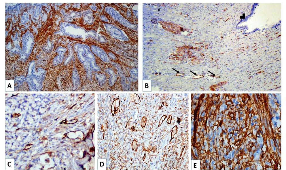
Background: Colorectal cancer is one of the most common cancers. Factors affecting survival include nodal positivity, lymphovascular invasion (LVI) and lymphangiogenesis (role in metastasis). Most endothelial markers stain lymphatics and blood vessels without discrimination. Recent marker D2-40 is specific for lymphatic endothelium.This study aims to interpret the utility of D2-40 in detecting LVI, which would be missed on H&E; and in analysing lymphatic vessel density and LVI as predictive markers for the risk of nodal metastases in colorectal carcinomas.Methods: Study was conducted on 40 specimens of colorectal carcinomas. Immunohistochemistry was performed with D2-40. Stastical analysis was by Spearman’s correlation and Chi square test Result: Mean age was 55.50 years. Of 40 cases, majority were in ascending colon followed by rectum, sigmoid, transverse and descending colon. 80.0% were adenocarcinomas, 15.0% mucinous adenocarcinomas and 5.0% signet-ring cell carcinomas. Tumors were moderately differentiated in 80.0% and poorly in 20.0%. Most patients were in stage T3 followed by T2 and T4. Most common nodal stage was N0 followed by N1, N2 and N3.LVI was detected in eleven more cases on D2-40 than H&E and correlated with lymphatic density and nodal status. Lymphatic density correlated with nodal status and pT. Peritumoral lymphatics were found in 95% cases and intratumoral lymphatics in 90%. Intratumoral lymphatics correlated with nodal status.Conclusion: D2-40 increased the detection rate of LVI as compared to H&E and showed the value of lymphangiogenesis in disease progression and metastasis. DOI: 10.21276/APALM.1122References
1. Parkin DM, Pisani P, Ferlay J. Global cancer statistics. CA Cancer J Clin. 1999;49(1):33-64.
2. Ferlay J, Shin HR, Bray F et al. Estimates of worldwide burden of cancer in 2008. Int J Cancer. 2010;127(12):2893–917.
3. Parums DV, Cordell JL, Micklem K et al. JC70: a new monoclonal antibody that detects vascular endothelium associated antigen on routinely processed tissue sections. J Clin Pathol. 1990;43:752-7.
4. Saad RS, Kordunsky L, Liu YL et al. Lymphatic microvessel density as prognostic marker in colorectal cancer. Mod Pathol. 2006;19:1317-23.
5. Marks A, Sutherland DR, Bailey D et al. Characterization and distribution of an oncofetal antigen (M2A antigen) expressed in testicular germ cell tumors. Br J Cancer. 1999;80:569-78.
6. Kahn HJ, Bailey D, Marks A. Monoclonal antibody, D2-40: A new marker of lymphatic endothelium, reacts with Kaposi's sarcoma and a subset of angiosarcomas. Mod Pathol. 2002;15:434-40.
7. Niakosari F, Kahn HJ, Marks A et al. Detection of lymphatic invasion in primary melanoma with monoclonal antibody D2-40: a new selective immunohistochemical marker of lymphatic endothelium. Arch Dermatol. 2005; 141:440-444
8. Maghraby HK, Elsarha AI, Saad RS. Peritumoral lymphatic vessel density as a prognostic parameter in endometrial carcinoma: An immunohistochemical Study. Indian J Pathol Microbiol. 2010;53(3):465-9
9. Stewart B, Wild C. Colorectal cancer. In: Bosman F, Hamilton S, Lambert R, editors. World cancer report. Lyon: IARC; 2014. P. 392-99
10. Compton CC. Pathology report in colon cancer : what is prognostically important? Dig Dis. 1999;17(2):67-79
11. Kahn HJ, Marks A. A new monoclonal antibody, D2-40, for detection of lymphatic invasion in primary tumors. Lab Invest. 2002;82(9):1255-7.
12. Fogt F, Zimmerman RL, Ross HM et al. Identification of lymphatic vessels in malignant, adenomatous and normal colonic mucosa using the novel immunostain D2-40. Oncol Rep 2004;11(1):47–50
13. Kawaura K, Fujii S, Murata Y et al. The Lymphatic Infiltration Identified by D2-40 Monoclonal Antibody Predicts Lymph Node Metastasis in Submucosal Invasive Colorectal Cancer. Pathobiology. 2007;74(6):328-35
14. Walgenbach-Bruenagel G, Tolba RH, Varnai AD et al. Detection of lymphatic invasion in early stage primary colorectal cancer with the monoclonal antibody D2-40. Eur Surg Res. 2006;38(5):438-44
15. Kuroyama S, Kobayashi N, Ohbu M et al. Enzyme histochemical analysis of lymphatic vessels in colon carcinoma: Occurrence of lymphangiogenesis within the tumor. Hepatogastroenterology. 2005;52(64):1057-61
16. Parr C, Jiang WG. Quanitative analysis of lymphangiogenic markers in human colorectal cancer. Int J Oncol. 2003;23(2):533–9
17. Naik VR, Jaafar H, Seng CE. Lymphatic channel density in colorectal adenocarcinoma. Indian J Pathol Microbiol. 2010;53(1):12-4.
18. Gombos Z, Xu X, Chu CS et al. Peritumoral lymphatic vessel density and vascular endothelial growth factor C expression in early stage squamous cell carcinoma of the uterine cervix. Clin Cancer Res. 2005;11(23):8367-71.
19. Sipos B, Klapper W, Kruse ML et al. Expression of lymphangiogenic factors and evidence of intratumoral lymphangiogenesis in pancreatic endocrine tumors. Am J Pathol. 2004;165(4):1187-97
20. Padera TP, Kadambi A, di Thomaso E et al. Lymphatic metastasis in the absence of functional intratumor lymphatics. Science. 2002;296(5574):1883-6.
Published
2017-04-07
Issue
Section
Original Article
Authors who publish with this journal agree to the following terms:
- Authors retain copyright and grant the journal right of first publication with the work simultaneously licensed under a Creative Commons Attribution License that allows others to share the work with an acknowledgement of the work's authorship and initial publication in this journal.
- Authors are able to enter into separate, additional contractual arrangements for the non-exclusive distribution of the journal's published version of the work (e.g., post it to an institutional repository or publish it in a book), with an acknowledgement of its initial publication in this journal.
- Authors are permitted and encouraged to post their work online (e.g., in institutional repositories or on their website) prior to and during the submission process, as it can lead to productive exchanges, as well as earlier and greater citation of published work (See The Effect of Open Access at http://opcit.eprints.org/oacitation-biblio.html).

