
Fast and inexpensive self-made tissue microarray for immunohistochemical and in situ hybridization studies: examples with bladder cancer
Keywords:
Tissue microarray, HER-2 Chromogen in situ hybridization, bladder cancer
Abstract
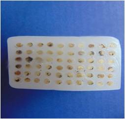
Background: The tissue microarray (TMA), first described by Battifora and first implemented in 1998 by Kononen et al, is a powerful research tool, allowing simultaneous analysis of specimens from a large number of cases on one slide by routine slides. Unfortunately, commercial aray instruments are very expensive and thus not suitable for laboratories with limited funds. We describe simple and cost-effective method for constructing of manual TMA that can be performed by any anatomic pathology laboratory, requiring minimum skill and time.Methods: Skin punch biopsy needle of 2-mm diameter was used for extracting cores from 15 positive control breast cancer cases, 15 normal bladder tissue and 30 muscle-invasive bladder cancer and for injecting into recipient block.Result: : We constructed TMA block using skin punch biopsy needle of 2-mm, and succesfulfully performed HER 2 immunodetection and Chromogen in situ hybridization studies, without substantial tissue loss. 10% of our tumor cases exhibited Her-2 neu overexpression. An open source software programme “ТМА-Ј“ was applied to facilitate management, viewing, analysis of tissue microarray images and associated clinico-pathology data.Conclusion: : This method could be done by any pathology laboratory and represents good and reliable alternative for commercially available, expensive devices and software solutions.DOI: 10.21276/apalm.2017.1166References
1. Battifora H. The multitumor (sausage) tissue block: novel method for immunohistochemical antibody testing.Lab Invest. 1986;55:244–248.
2. Kononen J, Bubendorf L, Kallioniemi A. Tissue microarrays for high-throughput molecular profiling of tumor specimens. Nat Med. 1998;4:844–47.
3. Wang H, Wang H, Zhang W, Fuller GN. Tissue microarrays: applications in neuropathology research, diagnosis, and education. Brain Pathol. 2002;12(1):95–107.
4. Bordeaux, J.M.; Cheng, H.; Welsh, A.W.; Haffty, B.G.; Lannin, D.R.; Wu, X.; Su, N.; Ma, X.J.; Luo, Y.; Rimm, D.L. Quantitative in situ measurement of estrogen receptor mRNA predicts response to tamoxifen. PLoS ONE 2012, 7, e36559.
5. Francis, GD, Jones MA, Beadle GF, Stein SR. Bright-field in situ hybridization for HER2 gene amplification in breast cancer using tissue microarrays: Correlation between chromogenic (CISH) and automated silver-enhanced (SISH) methods with patient outcome. Diagn. Mol. Pathol. 2009;18: 88–95.
6. Zlobec, I.; Terracciano, L.; Tornillo, L.; Gunthert, U.; Vuong, T.; Jass, J.R.; Lugli, A. Role of RHAMM within the hierarchy of well-established prognostic factors in colorectal cancer. Gut 2008, 57, 1413–1419.
7. Cheng, H.; Ballman, K.; Vassilakopoulou, M.; et al. EGFR expression is associated with decreased benefit from trastuzumab in the NCCTG N9831 (Alliance) trial. Br. J. Cancer 2014; 111:1065–1071
8. Schoenberg Fejzo M, Slamon DJ : Frozen tumor tissue microarray technology for analysis of tumor RNA, DNA, and proteins. Am J Pathol 159: 1645-1650,2001.
9. http://www.cap.org/apps/docs/committees/immunohistochemistry/summary_of_recommendations.pdf
10. Laé M, Couturier J, Oudard S, Radvanyi F, Beuzeboc P, Vieillefond A. Assessing HER2 gene amplification as a potential target for therapy in invasive urothelial bladder cancer with a standardized methodology: results in 1005 patients. Annals of Oncology. 2010;21: 815–819
11. Datta MW, Kahler A, Macias V, Brodzeller T, Kajdacsy-Balla A: A simple inexpensive method for the production of tissue microarrays from needle biopsy specimens: Examples with prostate cancer. Appl Immunohistochem Mol Morphol 2005;13: 96-103.
12. Shebl AM, Zalata KR, Amin MM, El-Hawary AK. An inexpensive method of small paraffin tissue microarrays using mechanical pencil tips. Diagn Pathol. 2011; 6:117
13. Wang SL, Yang CH, Chen HH, Chai CY. A simple and economical method for the manual construction of well-aligned tissue arrays. Pathol Res Pract. 2006; 202:485–486.
14. Pan CC, Chen PC, Chiang H. An easy method for manual construction of high-density tissue arrays. Appl Immunohistochem Mol Morphol. 2004; 12:370–372
15. Singh DK, Sakhuja P, Gondal R. Making and using inexpensive manually constructed tissue micro-array: experience of a tertiary care hospital in India. Indian J Pathol Microbiol. 2009; 52:304–309.
16. Pathak GS, Deshmukh SD, Ashturkar AV. Construction of tissue arrays without prefabricated recipient paraffin block experience of a novel technique in resource poor settings. Indian J Pathol Microbiol. 2011; 54:654–655.
17. Nocito A, Kononen J, Kallioniemi OP, Sauter G. Tissue microarrays (TMAs) for high-throughput molecular pathology research. Int J Cancer. 2001; 94:1–5.
18. Vogel UF, Bueltmann BD. Simple, inexpensive, and precise paraffin tissue microarrays constructed with a conventional microcompound table and a drill grinder. Am J Clin Pathol. 2006; 126:342–348.
19. Choi CH, Kim KH, Song JY, et al. Construction of High-Density Tissue Microarrays at Low Cost by Using Self-Made Manual Microarray Kits and Recipient Paraffin Blocks. Korean Journal of Pathology 2012; 46(6): 562-568.
Published
2017-02-19
Issue
Section
Original Article
Authors who publish with this journal agree to the following terms:
- Authors retain copyright and grant the journal right of first publication with the work simultaneously licensed under a Creative Commons Attribution License that allows others to share the work with an acknowledgement of the work's authorship and initial publication in this journal.
- Authors are able to enter into separate, additional contractual arrangements for the non-exclusive distribution of the journal's published version of the work (e.g., post it to an institutional repository or publish it in a book), with an acknowledgement of its initial publication in this journal.
- Authors are permitted and encouraged to post their work online (e.g., in institutional repositories or on their website) prior to and during the submission process, as it can lead to productive exchanges, as well as earlier and greater citation of published work (See The Effect of Open Access at http://opcit.eprints.org/oacitation-biblio.html).

