
A very rare occurrence of synchronous tumors of variable histology in bilateral ovaries
Keywords:
Synchronous, Ovarian Tumours, Immunohistochemistry
Abstract
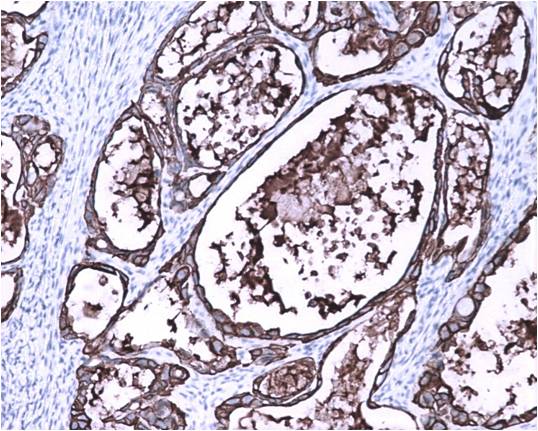
Introduction: Ovarian tumors make up 30% of all cancers of the female genital system. Surface epithelial tumors of the ovary are by far the commonest variety (90%) followed by germ cell tumors (30%) and sex cord stromal tumors (8%).We present a case of a 46 year old female with bilateral ovarian mass showing two distinct histopathological types of ovariantumorCase report: A 46 year old female came with complaints of irregular menstrual cycles, abdominal pain and distension since 8 months. Serum CA125 level was elevated. Ultrasonography revealed a complex cyst arising from the left adnexa. Patient underwent staging laparotomy. Left ovary sent for frozen section which was reported as malignant was a solid and cystic mass measuring 14x12x6 cm with the inner surface showing a solid area measuring 10x10x5 cm. Right ovary measured 5x4.4x4 cm with a 4x4x4cm grey white solid area seen on the cut surface. Microscopy revealed a surprising histopathological picture of a clear cell carcinoma of the left ovary and a granulosa cell tumor of the right ovary which was confirmed by immunohistochemistry. Both the tumors were confined to the ovaries without lymph node involvement.Conclusion: Morphologically different tumors arising from both ovaries is a rare occurrence with there being no case report of a synchronous clear cell carcinoma and granulosa cell tumordocumented in literature.References
1. Greenlee RT, Hill-Harmon MB, Murray T, Thun M. Cancer statistics, 2001. CA Cancer J Clin. 2001;51(1):15–36.
2. Levi F, Lucchini F, Negri E, Franceschi S. Cervical cancer mortality in young women in Europe: patterns and trends. Eur J [Internet]. 2000 [cited 2016 Aug 24]; Available from: http://www.sciencedirect.com/science/article/pii/S0959804900003464
3. Garg PP, Kerlikowske K, Subak L, Grady D. Hormone replacement therapy and the risk of epithelial ovarian carcinoma: a meta-analysis. ObstetGynecol. 1998;92(98):472–9.
4. Calle EE, Rodriguez C, Walker-Thurmond K, Thun MJ. Overweight, obesity, and mortality from cancer in a prospectively studied cohort of U.S. adults. N Engl J Med. 2003;348(17):1625–38.
5. Mungenast F, Thalhammer T. Estrogen biosynthesis and action in ovarian cancer. Front Endocrinol (Lausanne) [Internet]. 2014 [cited 2016 Aug 24];5(NOV). Available from: http://journal.frontiersin.org/article/10.3389/fendo.2014.00192/full
6. Walsh C, Holschneider C, Hoang Y, Tieu K. Coexisting ovarian malignancy in young women with endometrial cancer. Obstet. 2005;106:693-9
7. Saatli B, Yildirim N, Ozay AC, Koyuncuoglu M, Demirkan B, Saygili U. Synchronous tumors of the female genital tract: a 20-year experience in a single center. Ginekol Pol. 2014;85:441-5.
8. Preeti A, Arunachalam KA, Pradeep Y, Mati GM. Bilateral synchronous high-grade serous carcinoma and clear cell carcinoma in right and left ovaries with immunohistochemical confirmation: an exceptional finding. Indian J PatholMicrobiol. 2014;57:623-5
9. Sule A, Ochicha O, Yusuf I. Bilateral synchronous mature ovarian teratoma and mucinous cystadenoma. Arch IntSurg. 2015;5:174-6
10. Del Carmen MG, Birrer M, Schorge JO. Clear cell carcinoma of the ovary: A review of the literature. Gynecologic Oncology. 2012;126: 481–90.
11. Koukourakis G V, Kouloulias VE, Koukourakis MJ, Zacharias GA, Papadimitriou C, Mystakidou K, et al. Granulosa Cell Tumor of the Ovary: Tumor Review. Integr Cancer Ther . 2008;7:204–15.
12. Kanthan R, Senger J. The multifaceted granulosa cell tumours—myths and realities: a review. ISRN Obstet. 2012: 878635
13. Ayhan A, Salman MC, Velipasaoglu M, Sakinci M, Yuce K. Prognostic factors in adult granulosa cell tumors of the ovary : a retrospective analysis of 80 cases. J. 2009;20(3):158–63.
14. Otis CN. Protocol for the Examination of Specimens From Patients With Primary Tumors of the Ovary or Fallopian Tube Protocol applies to all primary borderline and malignant epithelial tumors. 2016
15. Mittal K, Soslow R. Application of immunohistochemistry to gynecologic pathology. Arch Pathol. 2008;132(3):402-23
Published
2016-12-10
Section
Case Report
Authors who publish with this journal agree to the following terms:
- Authors retain copyright and grant the journal right of first publication with the work simultaneously licensed under a Creative Commons Attribution License that allows others to share the work with an acknowledgement of the work's authorship and initial publication in this journal.
- Authors are able to enter into separate, additional contractual arrangements for the non-exclusive distribution of the journal's published version of the work (e.g., post it to an institutional repository or publish it in a book), with an acknowledgement of its initial publication in this journal.
- Authors are permitted and encouraged to post their work online (e.g., in institutional repositories or on their website) prior to and during the submission process, as it can lead to productive exchanges, as well as earlier and greater citation of published work (See The Effect of Open Access at http://opcit.eprints.org/oacitation-biblio.html).

