
Diagnostic Utility of AMCAR and p63 Cocktail Antibody in the Benign and Malignant Lesions of Prostate
DOI:
https://doi.org/10.21276/apalm.2800Keywords:
immunohistochemical analysis, prostate lesions, AMACR/p63 Cocktail antibodyAbstract
Background: Histopathological examination of prostatic specimen is gold standard for the diagnosis of prostate cancer. Current study evaluates the expression of AMACR and p63 in the prostate lesions using AMACR and p63 cocktail.
Materials and Method: Total of 180 cases were collected and Haematoxylin and Eosin staining performed followed by immunohistochemical analysis using AMACR and p63 antibody.
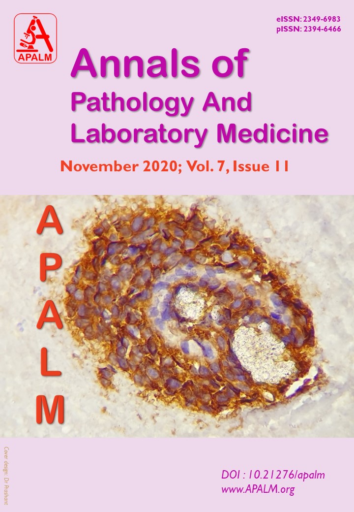
Result: Out of 180 cases, Benign Prostatic Hyperplasia is the most common lesion noted in about 120 cases. In this study, the predominant population was in the 6th to 7th decade of age. Most of the patients presented with difficulty in micturition. Immunohistochemistry revealed that p63 expression is positive in all normal basal cells, 118 cases (98.33%) were negative for AMACR and only 2 cases were showing focal and weak AMACR immunoreactivity. AMACR was positive in all the 6 HGPIN cases with variable intensity. Out of 5 LGPIN cases AMACR was positive in 3 cases with low intensity, remaining 2 cases shows AMACR negative. p63 is positive in all 11 PIN cases (LGPIN & HGPIN) showing discontinuous staining pattern. All 22 cases of Prostatic adenocarcinomas were negative for p63 and all cases expressed positive immunostaining with AMACR. A diagnosis of adenocarcinoma was made in 48% of atypical cases. Cases which were negative for both AMACR and p63 were diagnosed as Atypical Small Acinar Proliferation, for which further follow-up is required.
Conclusion: AMACR/p63 Cocktail antibody is very much useful as it saves time, tissue and is cost-effective.
References
2. Dabir PD, Ottosen P, Høyer S, Hamilton-Dutoit S. Comparative analysis of three-and two-antibody cocktails to AMACR and basal cell markers for the immunohistochemical diagnosis of prostate carcinoma. Diagnostic pathology. 2012 Dec;7(1):1-6.
3. Consolidated report of population based cancer registries 2001-2004: Incidence and distribution of cancer. Bangalore (IND): Coordinating Unit, National Cancer Registry Programme, Indian Council of Medical Research; 2006.
4. Kumaresan K, Kakkar N, Verma A, Mandal AK, Singh SK, Joshi K. Diagnostic utility of α-methylacyl CoA racemase (P504S) & HMWCK in morphologically difficult prostate cancer. Diagnostic Pathology. 2010 Dec 1;5(1):83.
5. Shah RB, Zhou M, LeBlanc M, Snyder M, Rubin MA. Comparison of the basal cell-specific markers, 34βE12 and p63, in the diagnosis of prostate cancer. Am J Surg Pathol. 2002 Sep 1;26(9):1161-8.
6. Signoretti S, Waltregny D, Dilks J, Isaac B, Lin D, Garraway L, Yang A, Montironi R, McKeon F, Loda M. p63 is a prostate basal cell marker and is required for prostate development. Am J Pathol. 2000 Dec 1;157(6):1769-75.
7. Parsons JK, Gage WR, Nelson WG, De Marzo AM. p63 protein expression is rare in prostate adenocarcinoma: implications for cancer diagnosis and carcinogenesis. Urology. 2001 Oct 1;58(4):619-24.
8. Beach R, Gown AM, De Peralta-Venturina MN, Folpe AL, Yaziji H, Salles PG, Grignon DJ, Fanger GR, Amin MB. P504S immunohistochemical detection in 405 prostatic specimens including 376 18-gauge needle biopsies. Am J Surg Pathol. 2002 Dec 1;26(12):1588-96.
9. Shah RB, Tadros Y, Brummell B, Zhou M. The diagnostic use of ERG in resolving an "atypical glands suspicious for cancer" diagnosis in prostate biopsies beyond that provided by basal cell and α-methylacyl-CoA-racemase markers. Human pathology. 2013 May 1;44(5):786-94.
10. Kuroda N. Application of combined immunohistochemical panel of AMACR (P504S)/p63 cocktail, cytokeratin 5 and D2-40 to atypical glands in prostatic needle biopsy. The Malaysian journal of pathology. 2014 Dec 1;36(3):169.
11. Xu J, Stolk JA, Zhang X, Silva SJ, Houghton RL, Matsumura M, Vedvick TS, Leslie KB, Badaro R, Reed SG. Identification of differentially expressed genes in human prostate cancer using subtraction and microarray. Cancer Res. 2000 Mar 15;60(6):1677-82.
12. Luo J, Zha S, Gage WR, Dunn TA, Hicks JL, Bennett CJ, Ewing CM, Platz EA, Ferdinandusse S, Wanders RJ, Trent JM. α-Methylacyl-CoA racemase: a new molecular marker for prostate cancer. Cancer Res. 2002 Apr 15;62(8):2220-6.
13. George E, Thomas S. A histopathologic survey of prostate disease in the sultanate of oman. Internet J Pathol 2005;3(2).
14. Barakzai MA, Mubarak M, Kazi JI. Histopathological lesions in transrectal ultrasound guided biopsies of prostate in patients with raised serum prostate specific antigen: a preliminary report. Nephro-Urol Mon 2011;3:186-90.
15. Anjorin AS, Adeniji KA, Ogunsulire IA. Histopathological study of prostatic lesions in Ilorin, Nigeria. Cent Afr J Med 1998;44:72-5.
16. Evans AJ. α-Methylacyl CoA racemase (P504S): overview and potential uses in diagnostic pathology as applied to prostate needle biopsies. Journal of clinical pathology. 2003 Dec 1;56(12):892-7.
17. Leav I, McNeal JE, Ho SM, Jiang Z. α-Methylacyl-CoA racemase (P504S) expression in evolving carcinomas within benign prostatic hyperplasia and in cancers of the transition zone. Human pathology. 2003;34(3):228-33.
18. Ananthanarayanan V, Deaton RJ, Yang XJ, Pins MR, Gann PH. Alpha"methylacyl"CoA racemase (AMACR) expression in normal prostatic glands and high"grade prostatic intraepithelial neoplasia (HGPIN): Association with diagnosis of prostate cancer. The Prostate. 2005 Jun 1;63(4):341-6.
19. Sanderson SO, Sebo TJ, Murphy LM, Neumann R, Slezak J, Cheville JC. An analysis of the p63/α-methylacyl coenzyme A racemase immunohistochemical cocktail stain in prostate needle biopsy specimens and tissue microarrays. American journal of clinical pathology. 2004 Feb 1;121(2):220-5.
20. Molinié V, Fromont G, Sibony M, Vieillefond A, Vassiliu V, Cochand-Priollet B, Hervé JM, Lebret T, Baglin AC. Diagnostic utility of a p63/α-methyl-CoA-racemase (p504s) cocktail in atypical foci in the prostate. Modern Pathology. 2004 Oct;17(10):1180-90.
21. Epstein JI, Herawi M. Prostate needle biopsies containing prostatic intraepithelial neoplasia or atypical foci suspicious for carcinoma: implications for patient care. The Journal of urology. 2006 Mar;175(3):820-34.
22. Sabata B, Babenko B, Monroe R, Srinivas C. Automated analysis of pin-4 stained prostate needle biopsies. InInternational Workshop on Prostate Cancer Imaging 2010 Sep 24 (pp. 89-100). Springer, Berlin, Heidelberg.
Downloads
Published
Issue
Section
License
Copyright (c) 2020 Sujitha Chougani, Sunandalakshmi G V, Durga Kharidehal, Ravi Sankar V, Santhi Vissa

This work is licensed under a Creative Commons Attribution 4.0 International License.
Authors who publish with this journal agree to the following terms:
- Authors retain copyright and grant the journal right of first publication with the work simultaneously licensed under a Creative Commons Attribution License that allows others to share the work with an acknowledgement of the work's authorship and initial publication in this journal.
- Authors are able to enter into separate, additional contractual arrangements for the non-exclusive distribution of the journal's published version of the work (e.g., post it to an institutional repository or publish it in a book), with an acknowledgement of its initial publication in this journal.
- Authors are permitted and encouraged to post their work online (e.g., in institutional repositories or on their website) prior to and during the submission process, as it can lead to productive exchanges, as well as earlier and greater citation of published work (See The Effect of Open Access at http://opcit.eprints.org/oacitation-biblio.html).
How to Cite


