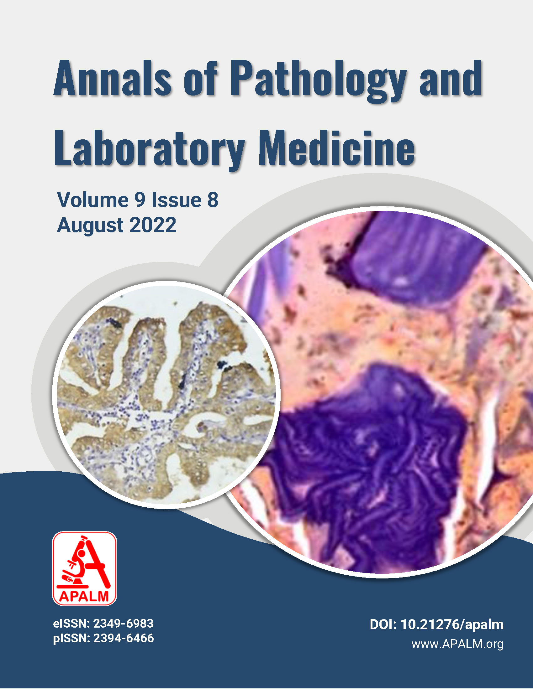
Diagnostic Utility of Galectin-3 in Papillary Lesions of Thyroid
DOI:
https://doi.org/10.21276/apalm.3189Keywords:
Galectin- 3, Immunohistochemistry, PTC, Papillary thyroid hyperplasiaAbstract
Introduction: Papillary thyroid carcinoma (PTC) is the most common malignant neoplasm of the neck. Most cases of PTC are diagnosed based on pathologic criteria. However, few thyroid lesions that mimic nuclear features or the architecture of PTC pose diagnostic problems. Papillary projections may be encountered in benign papillary hyperplasia of multinodular goiter, Hashimoto's thyroiditis, and Graves' disease. For this reason, the approach to these challenging lesions should include immunohistochemistry. Galectin-3(Gal-3) is an immunohistochemical marker that shows positivity in PTC. The present study was undertaken to investigate whether strong galectin-3 expression is an essential hallmark of PTC or papillary thyroid hyperplasia.
Material and Methods: Gal-3 expression was sought by immunohistochemistry in 33 cases of papillary patterns (on microscopy) of thyroid specimens received at our institution. The results obtained were statistically analysed.
Result and Conclusion: Of the 33 cases studied, 17 were PTC, and 16 were papillary hyperplasia. Immunohistochemical stain with Galectin -3 revealed a statistically significant P — value, which proves the tendency for Galectin -3 expression is higher in PTC than in papillary hyperplasia.
References
Htwe T T, Hamdi M M, Swethadri G K, Wong J O L, Soe M M, Abdullah M S. Incidence of thyroid malignancy among goitrous thyroid lesions from the Sarawak General Hospital 2000–2004. Singapore Med J 2009; 50:724-8.
Mary B. Casey, MD, Lohse, Lloyd RV. Distinction between Papillary Thyroid Hyperplasia and Papillary Thyroid Carcinoma by Immunohistochemical Staining for Cytokeratin 19, Gal-3, and HBME-1. Endocrine Pathology 2003; 14:55-60.
Sandra Fischer, Sylvia L. Asa. Application of Immunohistochemistry to thyroid neoplasms. Arch Pathol Lab Med 2008; 132:359-72.
Casey MB, Lohse CM, Lloyd RV. Distinction between papillary thyroid hyperplasia and papillary thyroid carcinoma by immunohistochemical staining for cytokeratin 19, galectin-3, and HBME-1. Endocrine pathology 2003; 14:55-60.
Baloch ZW, Livolsi VA. Pathology of Thyroid and Parathyroid Disease, In Mills SE editor. Sternberg’s Diagnostic Surgical Pathology. 6th ed. China: Wolters Kluwer; 2015.493-528.
Chan JKC. Tumors of the thyroid and parathyroid glands. In: Fletcher CDM (Ed). Diagnostic Histopathology of Tumors. 4th ed. Edinburgh, Scotland: Churchill Livingstone 2013. p.1177 -272.
Erickson LA, Yousef OM, Jin L, Lohse BS, Pankratz S, Lloyd RV. p27, kip1 Expression Distinguishes Papillary Hyperplasia in Graves’ Disease from Papillary Thyroid Carcinoma. Mod Pathol 2000; 13:1014–9.
Hirokawa M et al. Observer variation of encapsulated follicular lesions of the thyroid gland. Am J Surg Pathol 2002; 26:1508-14.
Franc B et al. Observer variation of lesions of the thyroid. Am J Surg Pathol 2003; 27:1177-9.
Fassina AS, Montesco MC, Ninfo V, Denti P, Masarotto G. Histopathological evaluation of thyroid carcinomas: reproducibility of the “WHO†classification. Tumors 1993; 79:314-20.
Heitz P, Maser H, Jacques Staub J. Thyroid cancer –A study of 573 thyroid tumors and 161 autopsy cases. Cancer 1967; 37:2329-37.
Shrikhande SS, Phadke AA. Papillary carcinoma of the thyroid gland. A clinicopathological study of 123 cases. Ind J. Cancer 1982; 19:87-92.
Sapio M R et al. Combined analysis of Gal-3 and BRAFV600E improves the accuracy of fine-needle aspiration biopsy with cytological findings suspicious for papillary thyroid carcinoma. Endocrine-Related Cancer 2007; 14:1089–97.
Beesley MF, McLaren KM. Cytokeratin 19 and Gal-3 immunohistochemistry in the differential diagnosis of solitary thyroid nodules. Histopathology 2002; 41:236–43.
Connie G, Chiu, Strugnell S S, Griffith O, Jones SJM, Gown AM, Blair Walker, Ivan R. Nabi, and Wiseman SM. Diagnostic Utility of Galectin -3 in Thyroid Cancer. The American Journal of Pathology 2010; 176:2067-81.
Park YJ et al. Diagnostic value of Gal-3. HBME-1, cytokeratin 19, high molecular weight cytokeratin, cyclin D1 and p27 (kip1) in the differential diagnosis of thyroid nodules. J Korean Med Sci 2007; 22:621–8.
Prasad ML, Pellegata NS, Huang Y, Nagaraja HN, Chapelle A, Kloos RT. Gal-3, fibronectin-1. CITED-1, HBME1 and cytokeratin-19 immunohistochemistry is useful for the differential diagnosis of thyroid tumors. Mod Pathol 2005; 18:48-57.
Barroeta JE, Baloch ZW, Lal P, Pasha TL, Zhang PJ, LiVolsi VA. Diagnostic value of differential expression of CK19, Gal-3, HBME-1, ERK, RET, and p16 in benign and malignant follicular- derived lesions of the thyroid: an immunohistochemical tissue microarray analysis. Endocr Pathol 2006; 17:225–34.
Coli A, Bigotti G, Zucchetti F, Negro F, Massi G. Gal-3, a marker of well-differentiated thyroid carcinoma, is expressed in thyroid nodules with cytological atypia. Histopathology 2002; 40:80–7.
Bartolazzi A, Gasbarri A, Papotti M, Bussolati G, Lucante T, Khan A. Application of an immunodiagnostic method for improving preoperative diagnosis of nodular thyroid lesions. Lancet 2001; 357:1644–50.
Weber KB, Shroyer KR, Heinz DE, Nawaz S, Said MS, Haugen BR: The use of a combination of Gal-3 and thyroid peroxidase for the diagnosis and prognosis of thyroid cancer. Am J Clin Pathol 2004; 122:524–31.
Downloads
Published
Issue
Section
License
Copyright (c) 2022 Raga Sruthi Dwarampudi, Yelikar B.R., Tejaswini Vallabha, Rodrigues Lynda D

This work is licensed under a Creative Commons Attribution 4.0 International License.
Authors who publish with this journal agree to the following terms:
- Authors retain copyright and grant the journal right of first publication with the work simultaneously licensed under a Creative Commons Attribution License that allows others to share the work with an acknowledgement of the work's authorship and initial publication in this journal.
- Authors are able to enter into separate, additional contractual arrangements for the non-exclusive distribution of the journal's published version of the work (e.g., post it to an institutional repository or publish it in a book), with an acknowledgement of its initial publication in this journal.
- Authors are permitted and encouraged to post their work online (e.g., in institutional repositories or on their website) prior to and during the submission process, as it can lead to productive exchanges, as well as earlier and greater citation of published work (See The Effect of Open Access at http://opcit.eprints.org/oacitation-biblio.html).


