
Histomorphological Study of Gestational Trophoblastic Lesions in a Tertiary Medical Centre: A Prospective Study
Keywords:
Gestational trophoblastic diseases, Complete mole, Partial mole, Invasive mole.
Abstract
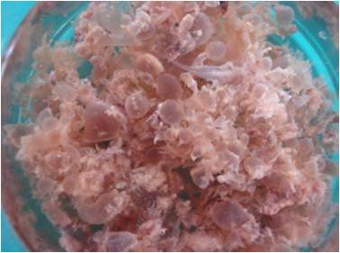
Background: Gestational trophoblastic diseases(GTDs) consists of pregnancy related disorders ranging from benign Hydatidiform mole, Invasive mole to neoplastic conditions including Choriocarcinoma, Placental site trophoblastic tumor and Epitheloid trophoblastic tumor with varying potential for local invasion and metastasis. GTDs mimic growth pattern encountered in early normal placental development, nonmolar abortions and variety of nontrophoblastic lesions. Therefore an appreciation of different types of GTDs with its histomorphological manifestations are important for the confirmation of diagnosis. Thus the study was undertaken. Materials & methods: The material for the study comprised of products of conception specimens received in the Department of Pathology, J.J.M. Medical college, Davangere, India during the period of 2 years. Detailed gross examination was done before fixation and subsequent microscopic analysis was done. Results: 65 cases of GTDs were diagnosed in 495 cases of products of conception. 37 cases of complete mole, 27 cases of partial mole and one case of Invasive mole were diagnosed. Diffuse hydropic swelling of the villi, round smooth villous outline, circumferential trophoblastic proliferation and focal hydropic swelling of the villi, irregular scalloping outline of the enlarged villi, focal trophoblastic cell proliferation were characteristic features of complete & partial mole respectively. Histomorphological features were analysed in all the cases and compared with studies done by others. Conclusion: The classification and histomorphological analysis of GTDs are important elements for better understanding of the disease. Studies are needed to look for ancillary markers in distinguishing different types of GTDs and for predicting the prognosis. DOI: 10.21276/APALM.1193References
1 Annie NY Chung. Gestational trophoblastic disease. In : Stanley JR, George LM, eds. Robboy’s pathology of female reproductive tract. Churchill Livginstone: Elsevier (Pub); 2009: 881-907.
2 Hertig AT. Human trophoblast coated by Charles B Hammond. Gestational trophoblastic neoplasia, history of current understanding. Obst & Gynec Clin of N-Am 1988;153:435-439.
3 Ie M S Michael TM, Robert JK. Gestational trophoblastic disease. In : Stacey EM, Jode KG, Harold AO, Victor R, Mart HS eds. Sternberg’s Diagnostic surgical pathology. Lippincot Williams and Wilkins (pub) 2010. pp2049-69
4 Liane D, Shirley GD, Donald PG. Gestational trophoblastic neoplasm. Morphology correlates of therapeutic response. Am J Obstet Gynec 1978;130(7):801-806.
5 Laurnence A. Cole. hCG, its free subunits and its metabolites role in pregnancy and trophoblastic disease. J Reprod Med 1998;43:3-10.
6 Michael JS, Neil SS, Ross SB. Gestational trophoblastic disease. Lancet 2010;376:717-729.
7 Michael JS, and Edward SN. Management of gestational trophoblastic disease. In: David MG, Gillian T, William PM, Martin G, Michael AQ eds. Gynecologic cancer . Churchill Livginstone: Elsevier (Pub); 2004:555-573.
8 George AH, Carl MH. Ectopic pregnancy. In: Larry JC eds. Text book of gynecology. W.B. Saunder’s Compnay; 1993 :242-260.
9 Ross SB, Donald PG. Gestational trophoblastic disease. In: Jonathan SB eds. Berek and Novak’s gynecology. Wolters Kluwer Health and Lippincott Williams and Wilkins 2006 pp.1581-1603
10 Rozina J, Rahat K, and Asmaa Q. Histopathological review of partial and complete hydatidiform moles in a tertiary care hospital, Lahore Pakistan, Biomedica 2011 June;27:76-80.
11 Fukunaga M, Shinichiro U and Yasuhiko E. Incidence of hydatidiform mole in a Tokyo Hospital : A 5 year (1989 to 1993) prospective, morphological and flow cytometric study. Human Pathol 1995;26(7):758-764.
12 Young HY, Hyun MK, Tchan KP, Chang KK and Yoo BL.Comparative cytogenetic and clinicopathologic studies on gestational trophoblastic neoplasia, especially hydatidiform mole. Yonsei Med J 1986;27(4):250-260.
13 Richard MC, Charles LH, Edwina JP, Henry JN, William F. Mc. Diagnostic considerations in molar gestations. Hum Pathol 1993;24(1);41-48.
14 Khairunnisa N, Guljareen H, Nizamuddin M, Ambreen H. Gestational trophoblastic disease: Experience at Nawabshah Hospital. J Ayub MedColl Abbottabad 2009;21(1):94-97.
15 Shakuntala C, Ambreen Q. Gestational trophoblastic neoplasms with special reference to invasive mole. Obstet Gynecol India 2007;57(2):124-127.
16 Amandeep KA, Shashi G, Vikram M, Rajat G, Rajni G. Invasive mole presenting as acute abdomen. J K Science 2011;13(1):35-36.
17 Debarmita M, Napur M, Ram PD, Ranu RB, Amiya KB, Subhash B.Partial invasive molar pregnancy – 2 case reports. Al Ameen J Med Sci 2010;3(1):91-93.
18 MiJK, KyuRK, JaeYR, JaniceML, HyangIL. Diagnostic and pathogenetic significance of increased stromal apoptosis and incomplete vasculogenesis in complete hydatidiform moles in very early pregnancy periods. Am J Surg Pathol 2006;30(3):362-69.
19 David K. Michael VZ, Terry H and Raymond WR. Very early complete hydatidiform mole. Hum Pathol 1996;27:708-713.
20 Kyu RK, Bong HP, Young OKH, Hyuck CK and Robboy SJ. The villous stromal constituents of complete hydatidiform mole differ histologically in very early pregnancy from the normally developing placenta. Am J Surg Pathol 2009;33:176-185.
21 Sebire.NJ.The diagnosis of gestational trophoblastic disease in early pregnancy: implications for screening, counseling and management Ultrasound Obstet Gynecol 2005; 25: 421–424.
22 Soper JT, Mutch DG, Schink JC. American College of Obstetricians and Gynecologists. Diagnosis and treatment of gestational trophoblastic disease: ACOG Practice Bulletin No. 53. Gynecol Oncol. 2004 Jun;93(3):575–85.
23 Overview of chemistry studies. In: Frances F, Marshall BD. A manual of laboratory and diagnostic tests. Lippincott Williams and Wilkins, and Wolters Kluwer Health (Pub) 2005 pp.338-482.
Published
2017-04-07
Issue
Section
Original Article
Authors who publish with this journal agree to the following terms:
- Authors retain copyright and grant the journal right of first publication with the work simultaneously licensed under a Creative Commons Attribution License that allows others to share the work with an acknowledgement of the work's authorship and initial publication in this journal.
- Authors are able to enter into separate, additional contractual arrangements for the non-exclusive distribution of the journal's published version of the work (e.g., post it to an institutional repository or publish it in a book), with an acknowledgement of its initial publication in this journal.
- Authors are permitted and encouraged to post their work online (e.g., in institutional repositories or on their website) prior to and during the submission process, as it can lead to productive exchanges, as well as earlier and greater citation of published work (See The Effect of Open Access at http://opcit.eprints.org/oacitation-biblio.html).

