
Prognostic significance of Ki 67 labelling index and P63 immunoreactivity in intra cranial and intra spinal meningiomas
Keywords:
Meningeal tumors, Meningiomas, Cytology, smear, Tumor grade, Ki67, P 63.
Abstract
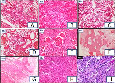
Background: Meningiomas are indolent tumors and their grading is based on histopathological parameters which have inherent limitations. Adjunct immunohistochemical markers are often used to overcome these limitations.Objective: To investigate the role of Ki67 and P63 immunoexpression in various grades of meningiomas.Methods: A retrospective analysis of 144 biopsies of intracranial and spinal meningothelial and nonmeningothelial tumors operated from January 2011 to May 2016 was carried out. Intraoperative squash smears were stained by 1% aqueous toluidine blue and rapid H&E method. Ki67 and p63 immunohistochemical staining was performed on paraffin sections of 125 histologically proven meningiomas cases. Grading was done according to the modified 2016 World Health Organization classification of CNS neoplasms .The results were analysed using statistical methods. Results: The total number of biopsy samples received was from144 cases. There was a female preponderance accounting for 68%of total cases. Positive cytohistological correlation was seen in 86.14% (125/144 cases). 112 out of 125 cases were found to be WHO grade I tumors out of which 20 showed a high Ki-67 LI and p63expression. 6 out of 7 cases of WHO Grade II meningiomas showed strong ki 67 and p63 nuclear expression whereas 3 anaplastic meningioma cases out of 6 WHO grade III meningiomas showed strong ki 67 and p63 nuclear positivity .Conclusion: The expression of P63 is variable in different grades of meningiomas. There was statistically significant increase of P63 protein expression and Ki 67 LI between the grades I to II and grades II to grade III. In the present study it was observed that 20/112 grade I meningiomas expressed high p 63 immunoreactivity contrary to other studies. High grade meningiomas showed increased Ki67 labelling index. P63 immunoexpression was high with predictability of recurrence across all the grades.References
1 Ostrom QT, Gittleman H, Farah P, Ondracek A, Chen Y, Wolinsky Y, et al. CBTRUS Statistical Report: Primary Brain and Central Nervous System Tumors Diagnosed in the United States 2006-2010. Neuro- Oncol. 2013; 15(2):ii1-ii56.
2 Kasuya H, Kubo O, Tanaka M, Amano K, Kato K, Hori T. Clinical and radiological features related to the growth potential of meningioma. Neurosurg Rev 2006; 29:293-7.
3 Lloyd RV, Kovacs K, Young Jr WF, Farrel WE, Asa SL, Trouillas J, Kontogeorgos G, Sano T, Scheithauer B, Horvath E. Tumours of the pituitary gland. In: DeLellis RA, Lloyd RV, Heitz PU, Eng C, editors.World Hearth Organization Classification of Tumours. Pathology and genetics of tumours of endocrine organs. Lyon: IARC Press; 2004. 9-47.
4 Karamitopoulou E, Perentes E, Melachrinou M, et al Proliferating cell nuclear antigen immunoreactivity in human central nervous system neoplasms. Acta Neuropathol 85:316-322, 1993
5 Nakasu S, Nakasu Y, Nakajima M, Matsuda M, Handa J. Preoperative identification of meningiomas that are highly likely to recur. J Neurosurg 1999; 90:455-62
6 Hahn HP, Bundock EA, Hornick JL Immunohistochemical staining for claudin-1 can help distinguish meningiomas from histologic mimics. Am J Clin Pathol. 2006 Feb; 125(2):203-8
7 Hopf NJ, Bremm J, Bohl J, Perneczky A. Image analysis of proliferating cells in tumors of the human nervous system: an immunohistological study with monoclonal Antibody Ki67. Neurosurg 1994; 35:917-23
8 LoMuzio L, Campisi G, Farina A. Effect of p63 expression on survival in oral squamous cell carcinoma. Cancer Invest. 2007; 25:466-469.
9 Sharifi N, Katebi M. An immunohistochemical study of p63 protein expression in meningioma. Iranian J Pathol 2008; 3: 146-150.
10 Devaprasath A, Chack G. Diagnostic validity of the Ki-67 labeling index using the MIB-1 monoclonal antibody in the grading of meningiomasNeurology India2003;51:336-340
11 Rushing EJ. Correlation of P63 immunoreactivity with tumor grade in meningiomas.Inter J Surg Pathol 2008; 16(1):38-42
12 Sharifi N, Katebi M. An immunohistochemical study of p63 protein expression in meningioma. Iranian J Pathol 2008; 3:146-150.
13 Jain D. Correlation of P63 protein expression with histological grade of meningiomas: An immunohistochemical study. Inter J Surg Pathol 2012; 20(4):349-354.
14 Guarnaschelli JJ, Stawicki SP. Brief communication: Recurrent brain meningiomas. OPUS 12 Scientist 2008; 2:32-4
Published
2016-11-08
Section
Original Article
Authors who publish with this journal agree to the following terms:
- Authors retain copyright and grant the journal right of first publication with the work simultaneously licensed under a Creative Commons Attribution License that allows others to share the work with an acknowledgement of the work's authorship and initial publication in this journal.
- Authors are able to enter into separate, additional contractual arrangements for the non-exclusive distribution of the journal's published version of the work (e.g., post it to an institutional repository or publish it in a book), with an acknowledgement of its initial publication in this journal.
- Authors are permitted and encouraged to post their work online (e.g., in institutional repositories or on their website) prior to and during the submission process, as it can lead to productive exchanges, as well as earlier and greater citation of published work (See The Effect of Open Access at http://opcit.eprints.org/oacitation-biblio.html).

