Histopathological Study of Villous Morphology in Spontaneous First Trimester Abortions
Keywords:
chorionic villi, fibrosis, vascularity, stromal fibrosis
Abstract
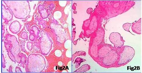
BACKGROUND: First trimester abortions are seen in 10 – 20 % of pregnancies. Sending the tissue evacuated after miscarriage for histopathological examination till date is a topic of debate as some professionals feel it is a waste of time and expensive, while on the other hand a school of thought still persists on doing the same.METHODS: We studied the various histopathological changes seen in the abortus tissue in first trimester spontaneous abortions over a period of one year from January to December 2015. A total of 100 slides and requests for histopathological examination of first trimester abortions were retrieved and studied in detail by two pathologists for the following features- Villous size, contour, vasculature, trophoblastic proliferation, perivillous and intervillous hemorrhage, perivillous fibrin deposition, stromal fibrosis, inflammation and decidual change. The observed changes were also categorized according to the weeks of abortion. Appropriate statistical tests were employed.RESULTS: Our study showed many dysmorphic features in the villi like reduced vessels per villous (72%), fibrosis (21.3%), hydropic change (32%) and abnormal trophoblastic proliferation (49%). Other features noted were villous hydrops, inter and perivillous fibrin deposition, inflammation, decidual change and Arias Stella reaction. Reduced vessels per villi and hydropic change were significantly associated with abortions happening at 8 - 10 weeks, while reduced patency of vessels, abnormal villous contour, ghost villi and trophoblastic proliferation were more associated with earlier dated abortions. CONCLUSION: Cases with dysmorphic features as seen in the present study are known to be associated with clinically significant conditions like diabetes, eclampsia and with certain chromosomal abnormalities. Such cases can not only be filtered for cytogenetic work up, but documentation of these features can also aid in counselling and planning of future pregnancies. Thus histopathological examination of abortus material is highly recommended.References
1.Crum, Christopher P. "The Female Genital Tract” In Ramzi S. Cotran, Vinay Kumar, and Tucker Collins: Robbins Pathologic Basis of Disease, , 8th ed. Philadelphia: W.B. Saunders, 2007:1079- 80.
2. Kalousek DK, Law AE. Pathology Of spontaneous abortion In Dimmick JE, Kalousek DK: Developmental Pathology of the embryo and Fetus: Phiiladelphia: JB Lippincot, 1992: 62-68.
3. Regan L, Clifford K. Sporadic and recurrent misccairage In Geofferry Chamberlain, Philip J Steer. Turnbull’s Obstetrics. 3rd edition Philadelphia: Churchill Livingstone 2001: 17 – 25.
4. Novak RF. A brief review of anatomy, Histology and ultrastructure of the full term placenta. Archives of Pathology and Laboratory Medicine 1991;115:654- 9.
5.Alsibiani SA. Value of Histopathologic Examination of Uterine Products after First-Trimester Miscarriage. BioMed Research International, 2014;1 :1- 5
6.Jauniaux E, Burton GJ. Pathophysiology of histological changes in early pregnancy loss. Placenta .2005;26:114-23.
7.Genest DR, Roberts D, Boyd T, Bieber FR. Fetoplacental histology as a predictor of karyotype: a controlled study of spontaneous first trimester abortions. Hum Pathol. 1995; 26: 201-9.
8. Ventura F, Rutigliani M, Bellini C, Bonsignore A, Fulcheri E. Clinical difficulties and forensic diagnosis: histopathological pitfalls of villous mesenchymal dysplasia in the third trimester causing foetal death. Forensic Sci Int. 2013;10: 229(1-3): e35–e41
9. Haque AU, Siddique S, Jafari MM, Hussain I, Siddiqui S. Pathology of Chorionic Villi in Spontaneous Abortions .International. Journal of pathology 2004;2: 5–9.
10. Hakvoort RA, Lisman BA, Boer K, Bleker OP, van Groningen K, van Wely M, Exalto N. Histological classification of chorionic villous vascularization in early pregnancy. Hum Reprod. 2006;1 :1291-4.
11.Karakaya YA, Ozer E. The role of Hofbauer cells on the pathogenesis of early pregnancy loss. Placenta. 2013;34: 1211-5.
12.Norris-Kirby A, Hagenkord JM, Kshirsagar MP, Ronnett BM, Murphy KM. Abnormal villous morphology associated with triple trisomy of paternal origin. J Mol Diagn 2010;12: 525-9.
13. Redline RW, Zaragoza M, Hassold T Prevalence of developmental and inflammatory lesions in non molar first-trimester spontaneous abortions. Hum Pathol. 1999; 30 :93-100.
14. Weber MA, Nikkels PG, Hamoen K, Duvekot JJ, de Krijger RR. Co-occurrence of massive perivillous fibrin deposition and chronic intervillositis: case report. Pediatr Dev Pathol. 2006; 9:234-8.
15.Fram KM. Histological analysis of the products of conception following first trimester abortion at Jordan University Hospital. Eur J Obstet Gynecol Reprod Biol. 2012;105:147-9.
16. Heath V, Chadwick V, Cooke I, Manek S, MacKenzie I Z. “Should tissue from pregnancy termination and uterine evacuation routinely be examined histologically?” British Journal of Obstetrics and Gynaecology 2000; 107:727–30.

Published
2016-11-07
Section
Original Article
Authors who publish with this journal agree to the following terms:
- Authors retain copyright and grant the journal right of first publication with the work simultaneously licensed under a Creative Commons Attribution License that allows others to share the work with an acknowledgement of the work's authorship and initial publication in this journal.
- Authors are able to enter into separate, additional contractual arrangements for the non-exclusive distribution of the journal's published version of the work (e.g., post it to an institutional repository or publish it in a book), with an acknowledgement of its initial publication in this journal.
- Authors are permitted and encouraged to post their work online (e.g., in institutional repositories or on their website) prior to and during the submission process, as it can lead to productive exchanges, as well as earlier and greater citation of published work (See The Effect of Open Access at http://opcit.eprints.org/oacitation-biblio.html).


