
A rare case of extraovarian granulosa cell tumor presenting as a retroperitoneal mass.
Keywords:
granulosa cell tumor, extraovarian, retroperitoneal
Abstract
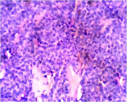
Introduction: Granulosa cell tumors (GCT) are the most common malignant sex cord—stromal tumors of the ovary. Rarely, they can occur at extraovarian site with very few cases mentioned in literature. Case history: A 45 yr old lady presented with pain abdomen and on ultrasonography, bilateral ovarian cyst was found. Patient underwent total abdominal hysterectomy with bilateral salpingoophorectomy and during surgery, a retroperitoneal mass was discovered which was excised. On gross examination, the mass measured 7x5x5cms, was solid with areas of hemorrhage and focal areas of necrosis . Microscopy showed solid sheets of cells with round to ovoid nuclei and scanty cytoplasm. Some of the cells showed nuclear grooves. A diagnosis of extraovarian adult granulose cell tumor was madeand immunostaining with inhibin was positive. Conclusion: Extraovarian granulosa cell tumor needs to be kept in mind in a female patient with a retroperitoneal mass. This case is presented due to its rarity and also to emphasise the fact that these tumors are often missed in the pre operative evaluation of patients and need histopathology for proper diagnosis.References
1. Geetha P, Nair MK. Granulosa cell tumours of the ovary. Aust N Z J Obstet Gynaecol 2010; 50: 216-20.
2. Schumer ST, Cannistra SA. Granulosa cell tumor of the ovary. J Clin Oncol. 2003;21(6): p. 1180-1189.
3. Kim SH, Park HJ, Linton JA, et al. Extraovarian granulosa cell tumour. Yonsei Med J. 2001;42(3): p. 360-360.
4. AG. B. Differentiation of mammalian embryonic gonad. Physiol Rev.. 1986;66(1): p. 71-117.
5. Motta PM, Makabe S. Germ cells in the ovarian surface during fetal development in humans. A three-dimensional microanatomical study by scanning and transmission electron microscopy. J Submicrosc Cytol. 1986; 81: p. 271-290.
6. P. C. Paul, J. Chakraborty, S. Chakrabarti, B. Chattopadhyay. Extraovarian granulosa cell tumor. Indian Journal of Pathology and Microbiology. 2009;52(2): p. 231-233.
7. M. Keitoku, I. Konishi, K. Nanbu, et al., “. Extraovarian sex cord-stromal tumor: case report and review of the literature. International Journal of Gynecological Pathology. 1997;16(2): p. 180-185.
8. Y. Sakai. Granulosa cell tumor arising in the wall of müllerian cyst of the broad ligament: report of a case and immunohistochemical study. Archives of Gynecology and Obstetrics. 2007;275(2): p. 145-148.
9. Young RH, Schully RE.. Sex cord-stromal, steroid cell, and other ovarian tumors with endocrine, and paracrine manifestations. In RJ K, editor. Blaustein's Pathology of the Female Genital Tract, 4th ed. New York: Springer-Verlag; 1995. p. 791-793
10. Manjiri R. Naniwadekar and N. J. Patil, “Extraovarian Granulosa Cell Tumor of Mesentery: A Case Report,”Pathology Research International, vol. 2010, Article ID 292606, 3 pages, 2010. doi:10.4061/2010/292606
11. Neeli, S. I. and P. R. Malur. (2010). “Primary Retroperitoneal Extraovarian Granulosa Cell Tumor: A Case Report.”UroToday Int J 3(6).
Published
2016-08-04
Issue
Section
Case Report
Authors who publish with this journal agree to the following terms:
- Authors retain copyright and grant the journal right of first publication with the work simultaneously licensed under a Creative Commons Attribution License that allows others to share the work with an acknowledgement of the work's authorship and initial publication in this journal.
- Authors are able to enter into separate, additional contractual arrangements for the non-exclusive distribution of the journal's published version of the work (e.g., post it to an institutional repository or publish it in a book), with an acknowledgement of its initial publication in this journal.
- Authors are permitted and encouraged to post their work online (e.g., in institutional repositories or on their website) prior to and during the submission process, as it can lead to productive exchanges, as well as earlier and greater citation of published work (See The Effect of Open Access at http://opcit.eprints.org/oacitation-biblio.html).

