
Clinicohistopathological Correlation in Leprosy Lesions: Study in a Tertiary Care Institute in Chhattisgarh
Abstract
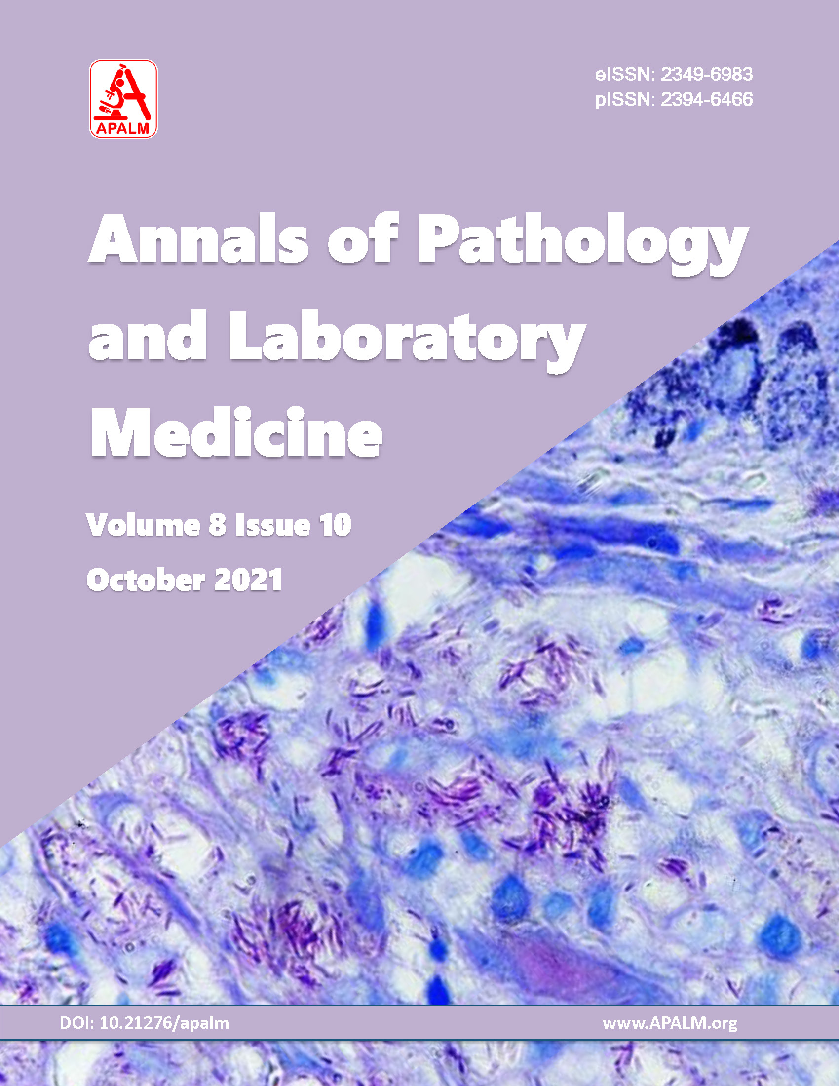
Background: Leprosy is a chronic granulomatous infectious disease, caused by Mycobacterium leprae. Early diagnosis is important to prevent morbidity caused by leprosy. However, accurate clinical or histopathological diagnosis is challenging especially in early skin lesions and in mid-borderline leprosy which shows overlapping features. Hence clinicohistopathological correlation is of utmost importance for accurate diagnosis. This study was conducted to evaluate the predominant subtype of leprosy in patients visiting for treatment in a tertiary care hospital, Chhattisgarh during the post-elimination phase in India. Methods: A retrospective study of 49 skin biopsies received from June 2019 to October 2020 was conducted in the Department of Pathology and Lab Medicine, AIIMS Raipur. Clinically suspected cases, outside diagnosed cases, and new lesions in treated cases of leprosy were included in this study. Clinical diagnosis was correlated with histopathological examination. Results: Among 49 skin biopsies, 34 cases were diagnosed as leprosy on histopathological examination. There was no evidence of leprosy in the remaining 15 cases. The most common type was borderline tuberculoid (35.3%), followed by Erythema nodosum leprosum (20.6%). Three cases were associated with type 1 lepra reaction. Among 34 confirmed cases of leprosy, 17(50.0%) cases were positive for acid-fast bacilli by Fite Faraco stain on histological sections. Maximum cases belonged to the age group of 31-40 years and the male to female ratio was 5.8:1. Conclusion: Histopathological examination of skin lesion along with Fite Faraco stain and clinical correlation is equally efficient for categorization and adequate treatment to achieve leprosy free world.References
Roy P, Dhar R, Patro P, Hoogar M.B, SahuS. Histopathological study of leprosy patients in a tertiary care hospital in Navi Mumbai. International journal of health sciences and research.IJHSR.2019;9(2).ISSN:2249-9571
Giridhar M, Arora G, Lajpal K, Singh Chahal K. Clinicohistopathological concordance in leprosy - a clinical, histopathological and bacteriological study of 100 cases. Indian J Lepr.2012;84(3):217-25.
Nadia S, Rashmi J, Sohaib A, Rawat SDS, Thamarai S, Meena H. clinicopathological correlation of leprosy: a 4 years retrospective study from a tertiary referral center in north India. Int J Med Res Health Sci. 2015;4(2):350-54.
Praba V, Narmadha C. Evaluation of Leprosy Cases in Correlation of Histopathology and Demonstration of Lepra Bacilli: A Prospective Study. Int J Sci Stud 2019;6(12):209-12.
Ramesh A, Sampath V, Shvedha M. A clinicopathological correlation in leprosy in a tertiary care teaching institution. Int J Res Dermatol. 2019;5(4):870-74.
Parkash O. Classification of leprosy into multibacillary and paucibacillary groups: an analysis. FEMS Immunol Med Microbio.2009;55(1):1-5.
Manandhar U, Adhikari RC, Sayami G. Clinico-histopathological correlation of skin biopsies in leprosy. J Pathol Nepal 2013;3:452-8.
Punia RPS, Dhingra H, Baliyan A et al. Clinicopathologic spectrum of Histoid leprosy. International Journal of Current Research. 2017;9(5):50765-769.
Vasaikar MS, Patil BM, Thakur RY. A Study of Histological Types of Leprosy Along with Clinico-Histopathological Correlation in a Tertiary Centre from North Maharashtra Region. Annals of Pathology and Laboratory Medicine. 2017;4(3):A321- A324Kalla G, Salodkar A, Kachhawa D. Clinical and histopathological correlation in leprosy. Int J Lepr Other Mycobact Dis. 2000 Jun;68(2):184-5.
Semwal S, Joshi D, Goel G, Asati D, Kapoor N. Clinico-histological correlation in Hansen's disease: Three-year experience at a newly established tertiary care center in central India. Indian J Dermatol 2018;63;465-8.
Joshi R. Clues to histopathological diagnosis of treated leprosy; Indian J Dermatol.2011;56(5): 505–9.
Veena S, Kumar P, Shashikala P, Gurubasavaraj H, Chandrasekhar HR, Murgesh. Significance of histopathology in leprosy patients with 1-5 skin lesions with relevance to therapy. J Lab Physicians 2011;3:21-4.
Cabic E, Cabic AG, Esposo SM, Dizon F, Quinones GJ, Guia A. Histopathological Detection of Mycobacterium Tuberculosis and Mycobacterium Leprae using a Modified Acid-Fast Technique. Phil Journal Path [Internet]. 2018Apr.17.
Ghosh R, Barua JK, Garg A, Barman BP. Dual infection with Mycobacterium tuberculosis and Mycobacterium leprae at the same site in an immunocompetent patient: An unusual presentation. Indian J Dermatol 2017;62:548.
Masuka JT, Mkhize Z, Pillay S, Mosam A. Concurrent pulmonary tuberculosis and lepromatous leprosy in a newly diagnosed HIV positive patient: a case report. BMC Pulm Med. 2021;21(1):207.
Copyright (c) 2021 Poonam Tulshiram Dambhare, Ashish Kumar Gupta, Nighat Hussain, Satyaki Ganguly

This work is licensed under a Creative Commons Attribution 4.0 International License.
Authors who publish with this journal agree to the following terms:
- Authors retain copyright and grant the journal right of first publication with the work simultaneously licensed under a Creative Commons Attribution License that allows others to share the work with an acknowledgement of the work's authorship and initial publication in this journal.
- Authors are able to enter into separate, additional contractual arrangements for the non-exclusive distribution of the journal's published version of the work (e.g., post it to an institutional repository or publish it in a book), with an acknowledgement of its initial publication in this journal.
- Authors are permitted and encouraged to post their work online (e.g., in institutional repositories or on their website) prior to and during the submission process, as it can lead to productive exchanges, as well as earlier and greater citation of published work (See The Effect of Open Access at http://opcit.eprints.org/oacitation-biblio.html).

