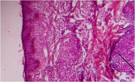
An evaluation of histopathological findings of skin biopsies in various skin disorders
Keywords:
Skin Biopsy, Histopathology, Leprosy,
Abstract
Background - Skin conditions are among the most common health problems in India. Skin biopsy is most common diagnostic technique for diagnosing skin disease. The interpretation of a skin biopsy requires clinicopathological correlation.Methods: The present study was undertaken in the Department of Pathology, M.G.M Medical College and M.Y. Hospital, Indore (M.P) to determine the incidence and age-sex distribution of various skin diseases, to study the various histopathological changes encountered in the course of the study and to establish clinic pathological correlation. A total number of 262 biopsies retrieved from the archives during the period of January 2007 to June 2008 along with skin biopsies sent for histopathological evaluation during June 2008 – June 2010 were included in the study. On the basis of histopathological classification the skin disorders are divided into eight groups.Result: Maximum number of cases belonged to group V disorders i.e. disorders showing perivascular, diffuse and granulomatous infiltrates of the reticular dermis 65(24.8 %), followed by group VI disorders that is tumors and cysts of the dermis 28(10.7 %). Out of 262 skin biopsies which came for Histopathological evaluation, 137 (52.3 %) cases were given definite diagnosis by microscopic examination of slides. Amongst 216 cases, clinico-histopathological correlation was seen in 95(44 %) cases. Highest number of cases 68(25.95 %) were observed in the age group of 21-30 years, with majority of the cases were male 149(56.9 %) while 113(43.1 %) cases were female.Conclusion: Leprosy is still most prevalent, thereby emphasizing stronger measures to control it.References
1. Grover S, Ranyal RK, Bedi MK. A cross section of skin diseases in rural Allahabad. Ind J Derm2008;53:179-81.
2. Elder DE et al in Elder DE, Editor in chief. Lever’s Histopathology of the skin. 10th ed. Lippincott Williams and Wilkins; 2009. p 103-132.
3. Das S, Chatterjee T. Pattern of skin diseases in a peripheral hospital’s skin OPD. Ind J Derm2007; 52:93-5.
4. Das KK. Pattern of dermatological diseases in Gauhati Medical college and hospital. Ind J Derm Venereo Leprol 2003;69:16-18.
5. Rao GS, Kumar SS, Sandhya. Pattern of skin diseases in an Indian village. Ind J Med Sci2003; 57:108-10.
6. Veena S, Kumar P, Shashikala P, Gurubasavaraj H, Chandrashekhar HR, Murugesh. Significance of Histopathology in leprosy patients with 1-5 skin lesions with relevance to therapy. J Lab Physicians 2011: 3:21-4.
7. Grace DF, Bendale KA, Patil YV. Spectrum of pediatric skin biopsies. Indian J Dermatol 2007;52:111-5.
8. Thapa DM. Textbook of Dermatology, Venere-ology & Leprology.3rd edition Elsevier 2009
9. Mysore V. Fundamentals of pathology of skin .3rd edition. B.I Publications 2008
10. WHO, weekly epidemiological record, no 35, 27th Aug 2010.
11. Beliaeva TL. The population incidence of warts. Vestn Dermatol Venerol. 1990;2:55-8.
12. Kruger C, Schallreuter K U. A review of worldvide prevalence of vitiligo in children/adolescents & adults. Int J Derma 2012
13. Parisi R, Symmons DP, Griffiths CE, Ashcroft DM. Global epidemiology of psoriasis: a sys-tematic review of incidence and prevalence. J Invest Dermatol. 2013;133:377-85.

Published
06-02-2015
Issue
Section
Original Article
Authors who publish with this journal agree to the following terms:
- Authors retain copyright and grant the journal right of first publication with the work simultaneously licensed under a Creative Commons Attribution License that allows others to share the work with an acknowledgement of the work's authorship and initial publication in this journal.
- Authors are able to enter into separate, additional contractual arrangements for the non-exclusive distribution of the journal's published version of the work (e.g., post it to an institutional repository or publish it in a book), with an acknowledgement of its initial publication in this journal.
- Authors are permitted and encouraged to post their work online (e.g., in institutional repositories or on their website) prior to and during the submission process, as it can lead to productive exchanges, as well as earlier and greater citation of published work (See The Effect of Open Access at http://opcit.eprints.org/oacitation-biblio.html).


